Gülfize Coşkun¹, Veysel Olgun¹, Fikret Dirilenoğlu², Fadime Gül Salman³, Murat Oktay³, Serdar Balcı³, İlknur Türkmen³
¹Technomind Dijital Sistemler, BÜDOTEK Teknopark, Istanbul, Türkiye
²Yakın Doğu Üniversitesi Tıp Fakültesi, Tıbbi Patoloji Anabilim Dalı, Lefkoşa, Kıbrıs
³Memorial Hospitals Group, Department of Pathology, Istanbul, Turkey
Introduction:
Digital pathology leverages advanced image analysis techniques to quantify Ki-67 expression in a more objective and reproducible manner. Computer-assisted image analysis tools aid pathologists in efficiently analyzing large datasets, leading to more accurate and standardized results. However WSIs are huge in size while containing inherent information. Compressing the images may give rise to information loss. Here, we tried to assess the outcome of image compression on the Ki-67 proliferation index.

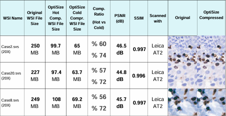
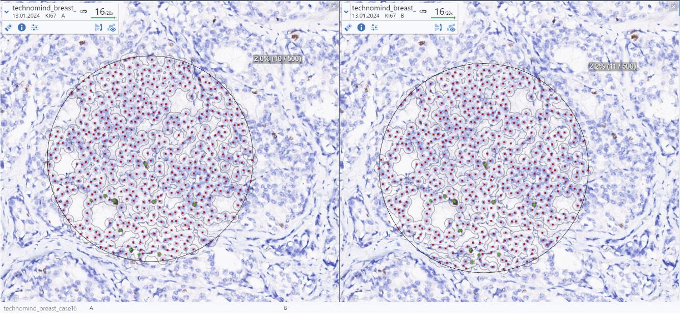
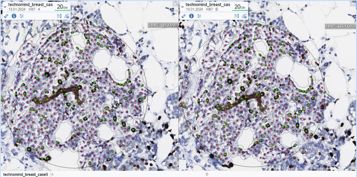
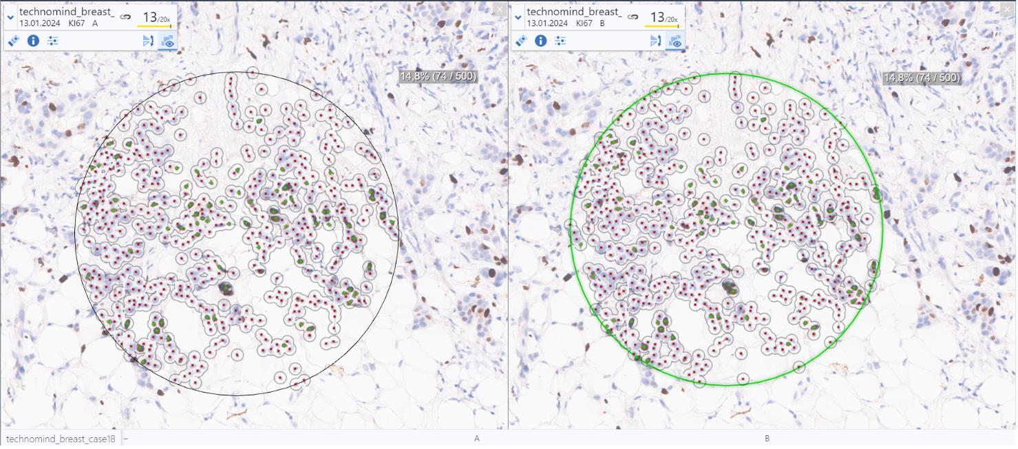
Materials and Methods: Twenty breast cases with WSIs were included. Original images were exported from Sectra in .svs format. Using OptiSize Technomind software, the images were compressed into .tiff format. The compressed images and original images were reimported into Sectra. The original image and the image compressed by OptiSize were randomly placed in the Sectra application for pathologists to review. Pathologists did not know which image was the original and which was the compressed image. First, they examined the images themselves and then used the Ki-67 test available in the Sectra application. Both images were aligned and synchronized to select the same areas. Three corresponding areas were marked to measure the Ki-67 proliferation index, using Sectra’s built-in tool. Furthermore, an assessment was conducted on the mathematical analyses, specifically the Peak Signal-to-Noise Ratio (PSNR) and Structure Similarity Index Measure (SSIM), applied to both the original and compressed images.

Results:
The findings indicate a notable compression rate of 59% and median Ki-67 index was 17.9% in original images (min: 0.1%, max: 54.2%), and 18.2% in compressed (min: 0.1%, max: 55.1%) (p= 0.07). Individual differences are detected without any predilection towards a group. The mathematical verification of the image compression ratio performed a promising outcome, characterized by a PSNR value of 44.20dB and an SSIM value of 0.972. In evaluations by expert pathologists, OptiSize performed remarkably well. The expert pathologists examined the original and OptiSize compressed images without knowing which image was the original and found both images to be of good quality with no visible differences between them. In the future, compressed images will be analyzed using additional AI-based tools for Ki-67, and corresponding index values will be examined. OptiSize is compatible with many kind of file format .svs .mrxs .tiff .svslide .ndpi .vms .scn and also .ome.tif.
